

Tuning the Brain with Electricity, Magnetism, and Sound
June 12, 2025
In The Brain's Operating System, we looked at how the brain's neural circuits work: how neurons fire, how excitation and inhibition stay in balance, and how networks of neurons underlie everything the brain does. This post asks the obvious next question: when those circuits malfunction, how can we deliberately influence them?
Decades of work on neuromodulation have produced several ways of delivering energy to the brain. Electrical currents, magnetic fields, and sound waves each interact with neural tissue through a different physical mechanism, and those differences go a long way toward explaining which approach suits which condition.
The Physics of Influencing Neurons
All brain stimulation methods share one goal: changing the electrical state of neurons. Every neuron maintains a voltage across its membrane (the resting potential covered in that post), and stimulation techniques work by manipulating that electrical environment.
Ion channels are the lever. These membrane proteins control the flow of charged particles such as sodium, potassium, and calcium into and out of the cell. When enough positive charge enters a neuron to push it past threshold, it fires an action potential: sodium rushing in triggers the spike, and potassium flowing out resets the cell.
External energy can influence this process in two ways. It can nudge neurons closer to or further from their firing threshold without actually making them fire (subthreshold effects), or it can deliver enough energy to trigger action potentials directly (suprathreshold effects). This distinction separates the gentle modulators from the direct drivers.
Electrical Stimulation
Applying electrical current is the most direct way to change the voltage environment around neurons. The three main electrical approaches, however, differ enormously in precision and invasiveness.
Transcranial Direct Current Stimulation (tDCS): Broad Network Modulation
tDCS applies a weak current, typically 1 to 2 milliamps, through sponge or gel electrodes placed on the scalp. Each electrode is usually about the size of a playing card and soaked in saline, connected to a battery-powered device roughly the size of a smartphone. A couple of milliamps is a small current to begin with, and only a fraction of it reaches the brain: after passing through the scalp and skull, the resulting electric field shifts neurons' membrane voltage by just 0.1 to 0.5 millivolts, far below what is needed to trigger an action potential.
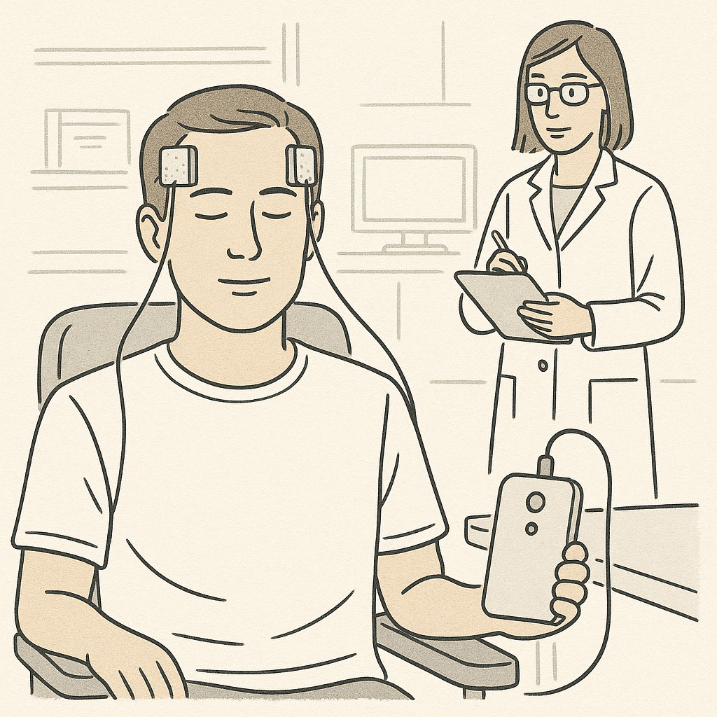
Transcranial direct current stimulation (tDCS) uses electrodes on the scalp to deliver weak electrical current, gently shifting neural excitability across broad brain regions.
tDCS therefore works entirely through subthreshold modulation. It doesn't force neurons to fire; it shifts their resting potential so that they respond a little more, or a little less, readily to their normal synaptic inputs. It's like adjusting the sensitivity of a smoke detector: the alarm isn't being set off, but the threshold for triggering it has changed.
Because the current spreads diffusely through tissue, tDCS influences broad areas of cortex rather than any precise target. Its effects are correspondingly modest and vary considerably between individuals, which is why it is mostly explored for conditions where a gentle shift in the excitability of a whole network might help.
Deep Brain Stimulation (DBS): Surgical Precision
DBS sits at the opposite extreme. A thin electrode, about 1.3 mm in diameter, is surgically implanted into a specific brain target and connected by a cable tunneled under the skin to a pulse generator, similar to a cardiac pacemaker, implanted below the collarbone.

Deep brain stimulation (DBS) delivers precise electrical pulses through a surgically implanted electrode connected to a pulse generator in the chest.
Once activated, the system delivers continuous high-frequency electrical pulses (typically 130 to 185 Hz), with settings adjusted via an external programmer. Its precision comes from proximity: the electrode contacts sit just millimeters from the target neurons, so stimulation can be confined to a specific neural population.
In Parkinson's disease, that precision matters. DBS targets specific nuclei within the brain's motor circuits, and the high-frequency pulses disrupt pathological synchrony, the harmful lockstep firing that emerges in these circuits when dopamine is lost. The cost is neurosurgery, with the risks and expense that implies.
Cortical Electrode Arrays: Cellular-Level Interface
The finest electrical control comes from electrode arrays placed directly on or in brain tissue. These take several forms: flexible grids that conform to the brain's surface, rigid arrays of needle-like microelectrodes, or thin strips that slide into the folds of the cortex. A typical grid carries 64 to 256 contacts, each from under a millimeter to a few millimeters across.
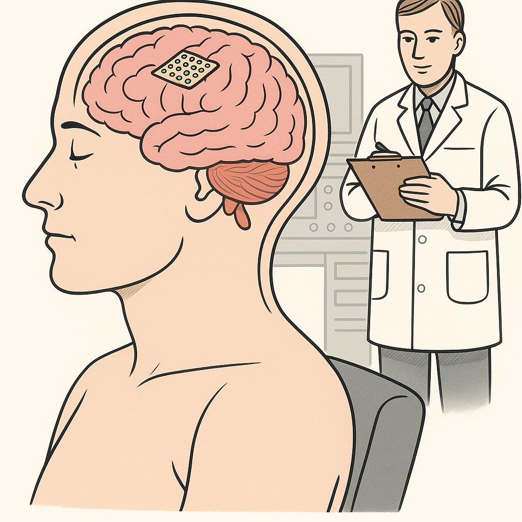
Electrode arrays placed directly on brain tissue enable precise monitoring and stimulation of small groups of neurons.
Because the electrodes touch the tissue directly, very small currents are enough, and each contact may influence only a few hundred neurons. This makes arrays a powerful tool for researchers and clinicians probing the function of specific circuits, though like DBS they require surgery.
Magnetic Stimulation: Induction at a Distance
Transcranial magnetic stimulation (TMS) takes a different route to the same end. Rather than applying current directly, it uses rapidly changing magnetic fields to induce electrical currents inside brain tissue, exploiting Faraday's law of electromagnetic induction.
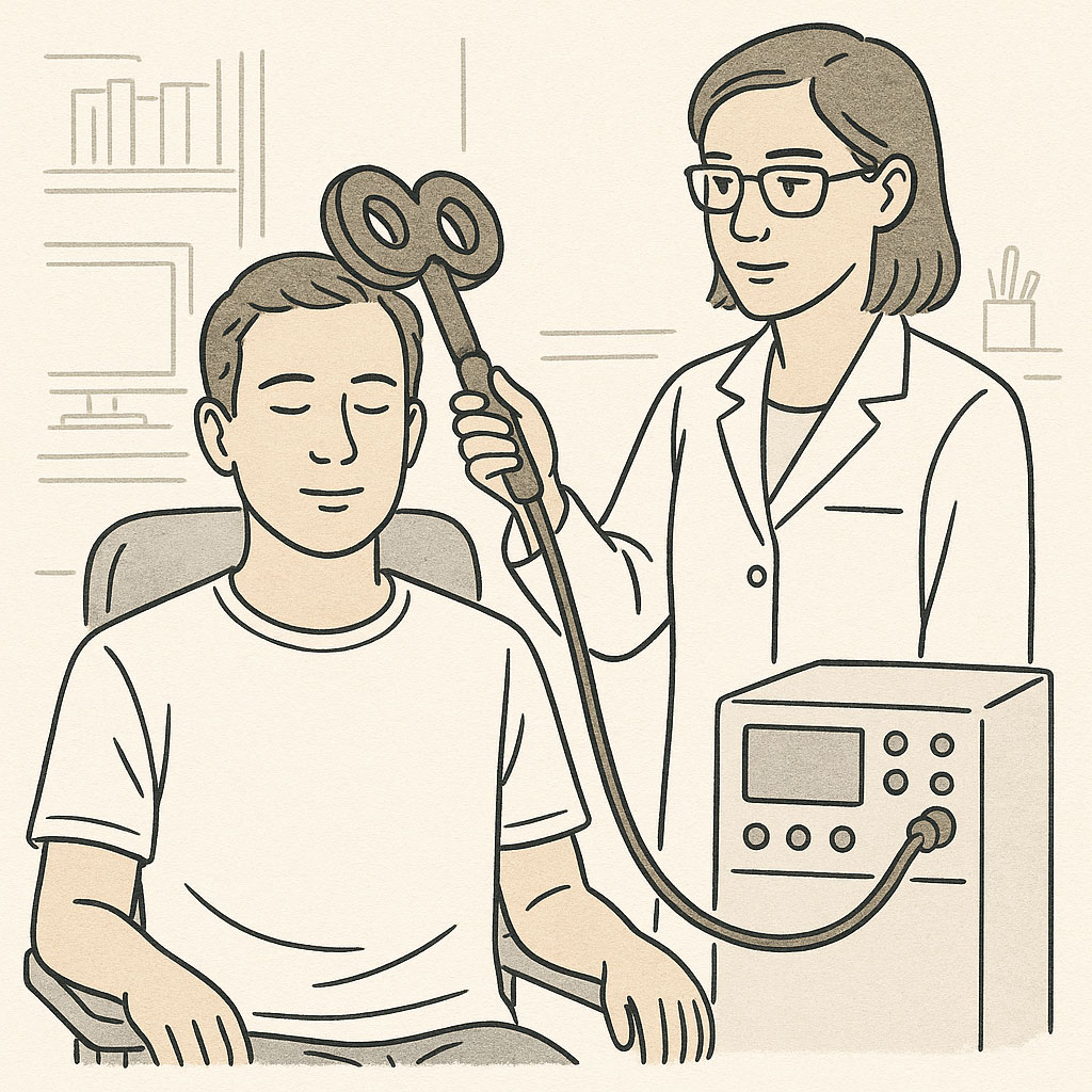
Transcranial magnetic stimulation (TMS) uses a figure-8 shaped magnetic coil positioned against the scalp to induce electrical currents in targeted brain regions.
In a TMS session, a magnetic coil, usually shaped like a figure-8 and about the size of a small dinner plate, is held against the scalp. Each brief, powerful magnetic pulse passes through the skull unimpeded and induces circular currents in the tissue beneath, accompanied by a sharp click from the coil. The induced currents can be tuned either to trigger action potentials outright or to subtly shift excitability, though most therapeutic protocols use suprathreshold intensities.
The same physics sets TMS's main limit. While the magnetic field crosses the skull easily, the induced currents weaken rapidly with distance from the coil, so TMS mainly affects the outer 2 to 3 centimeters of the brain, like lighting up the top floors of a building while the basement stays dark. Even so, it has found real success in treating depression, where stimulating specific cortical areas influences the deeper mood-regulation networks they connect to. Its precisely timed pulses have also made TMS a valuable research tool for mapping brain function, by briefly disrupting one area and observing what changes.
Ultrasound Stimulation: Mechanical Modulation
Transcranial ultrasound stimulation is the newest of these technologies, and the one we work on at NeuroHarmonics. It uses focused sound waves at frequencies far above human hearing, and its mechanism differs in kind from the electrical and magnetic approaches.
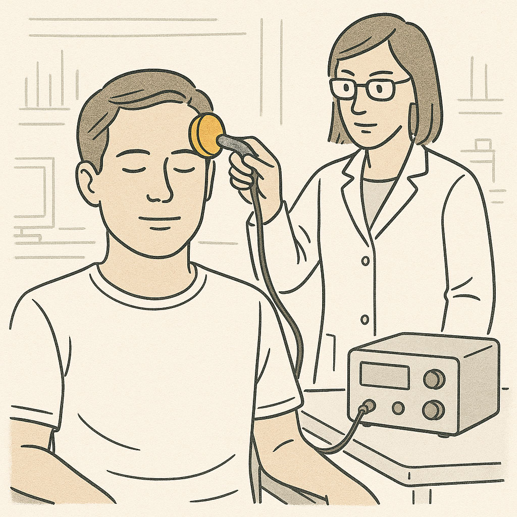
Focused ultrasound uses high-frequency sound waves to mechanically influence neural activity with millimeter precision, even in deep brain structures.
Rather than manipulating electrical fields, ultrasound delivers mechanical pressure waves that minutely deform neural tissue many thousands of times per second. This mechanical stimulation is thought to act on mechanosensitive ion channels, proteins that open or close in response to physical force, and so change the neuron's electrical state. The exact cellular and circuit-level mechanisms are still being worked out.
What sets ultrasound apart is focusing. Sound waves can be focused far more tightly than electrical or magnetic fields, down to volumes a few millimeters across, and they can be focused deep inside the brain rather than only near its surface. No other non-invasive method combines that depth and precision, which is why we believe ultrasound can reach targets that have so far required surgery. We described how we built and tested a system that does exactly this in Our Journey. Clinical validation is still ahead, but the physics is what drew us to the approach.
Comparing the Approaches
Each modality trades off precision, depth, and invasiveness differently:
| Method | Energy Type | Precision | Depth Reach | Invasiveness | Primary Mechanism |
|---|---|---|---|---|---|
| tDCS | Electrical | Low | Superficial | Non-invasive | Subthreshold |
| TMS | Magnetic | Moderate | Superficial (2-3 cm) | Non-invasive | Typically suprathreshold |
| DBS | Electrical | High | Deep | Invasive (surgery) | Suprathreshold |
| Ultrasound | Acoustic | High | Deep | Non-invasive | Subthreshold (mechanical) |
| Electrode Arrays | Electrical | Very high | Cortical surface | Invasive (surgery) | Suprathreshold |
The table makes the field's long-standing trade-off plain: precision has historically required surgery, while non-invasive methods have been blunt instruments. Focused ultrasound is the most direct attempt to break that trade-off, offering surgical-level targeting without an incision.
Matching the Tool to the Problem
Choosing a stimulation technology means matching the tool to the problem. Some disorders may respond to broad network modulation; others hinge on hitting a millimeter-scale target. And precision is only one dimension. The timing and pattern of stimulation matter too: whether energy arrives as continuous pulses or as bursts that mimic natural brain rhythms helps determine whether effects last minutes or months.
Our own work is a bet on that combination of depth and precision. You can see the system we're building in A First Look at Our Neuromodulation Platform.